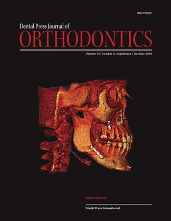
The evolution of imaging diagnostics for Orthodontics
In the 1970s, the electronic technologiesdeployed for space exploration launched averitable revolution in imaging diagnosticscapacity—especially in the field of computertomography. It is curious to note that before wewere able to delve deeper into the human bodywe had to first travel into...
Autores: Jorge Faber,
messages.RevistaAutores:
LEIA MAIS
Digital impressions and handling of digital models: The future of Dentistry
New digital impression methods are currentlyavailable in the market, and soon thelong-awaited dream of sparing patients one ofthe most unpleasant experiences in dental clinics,the taking of dental impressions, will be replacedby intraoral digital scanning.Both in orthodontics and restorative...
Autores: Waldemar D. Polido,
messages.RevistaAutores:
LEIA MAIS
Orthodontic traction: possible effects on maxillary canines and adjacent teeth
Professionals who resist and restrict the indicationof orthodontic traction, especially caninetraction, often justify their stance by citing thefollowing reasons:1) Lateral Root Resorption in lateral incisorsand premolars.2) External Cervical Resorption of caninesdue to canine traction.3)...
Autores: Alberto Consolaro,
messages.RevistaAutores:
LEIA MAIS
An interview with Lucia Helena Soares Cevidanes
It gives me great pleasure to conduct an interview with Professor Lucia Cevidanes, an example of humbleness, courageand determination. Born in Caratinga, Minas Gerais, she attended dentistry at the Federal University of Goiás and earneda Masters Degree in Orthodontics at UMESP, where she was...
Autores: Ary Dos Santos-Pinto, Daniela Gamba Garib, Alexandre Trindade Motta, Liliana Maltagliati,
messages.RevistaAutores:
LEIA MAIS
Analysis of initial movement of maxillary molars submitted to extraoral forces: a 3D study
Objective: To analyze maxillary molar displacement by applying three different angulationsto the outer bow of cervical-pull headgear, using the finite element method (FEM).Methods: Maxilla, teeth set up in Class II malocclusion and equipment were modeledthrough variational formulation and their...
Autores: Antônio Carlos De Oliveira Ruellas, Eduardo Franzotti Sant’anna, Janaina Cristina Gomes, Mônica Tirre De Souza Araújo, Carlos Nelson Elias, Luciana Rougemont Squeff, Norman Duque Penedo, Giovana Rembowski Casaccia, Jayme Pereira Gouvêa,
messages.RevistaAutores: Tooth movement, Finite element method, Headgear,
LEIA MAIS
2D / 3D Cone-Beam CT images or conventional radiography: Which is more reliable?
Objective: To compare the reliability of two different methods used for viewing and identifyingcephalometric landmarks, i.e., (a) using conventional cephalometric radiographs,and (b) using 2D and 3D images generated by Cone-Beam Computed Tomography. Methods:The material consisted of lateral view 2D...
Autores: Oswaldo De Vasconcellos Vilella, Carolina Perez Couceiro,
messages.RevistaAutores: cone-beam computed tomography, Orthodontics, Radiography,
LEIA MAIS
Evaluation of referential dosages obtained by Cone-Beam Computed Tomography examinations acquired with different voxel sizes
Objectives: The aim of this study was to evaluate the dose–area product (DAP) andthe skin entrance dose (SED), using protocols with different voxel sizes, obtained withi-CAT Cone-Beam Computed Tomography (CBCT), to determine the best parametersbased on radioprotection principles. Methods: A...
Autores: Paulo Sérgio Flores Campos, Marianna Guanaes Gomes Torres, Marcus Navarro, Iêda Crusoé-Rebello, Nilson Pena Neto Segundo, Marlos Ribeiro,
messages.RevistaAutores: voxel, cone-beam computed tomography, Radiation,
LEIA MAIS
Linear measurements of human permanent dental development stages using Cone-Beam Computed Tomography: A preliminary study
Objective: To determine the linear measurements of human permanent dentition developmentstages using Cone-Beam Computed Tomography. Methods: This study wasbased on databases of private radiology clinics involving 18 patients (13 male and 5 female,with age ranging from 3 to 20 years). Cone-Beam...
Autores: Carlos Estrela, José Valladares Neto, Mike Reis Bueno, Orlando Aguirre Guedes, Olavo Cesar Lyra Porto, Jesus Djalma Pécora,
messages.RevistaAutores: cone-beam computed tomography, Computed tomography, Incomplete root formation, Tooth development, Apexogenesis,
LEIA MAIS
Skeletal displacements following mandibular advancement surgery: 3D quantitative assessment
Objective: To evaluate changes in the position and remodeling of the mandibular rami,condyles and chin with mandibular advancement surgery through the superimposition of3D Cone-Beam Computed Tomography (CBCT) models. Methods: This prospective observationalstudy used pre-surgery and post-surgery...
Autores: Marco Antonio De Oliveira Almeida, Felipe De Assis Ribeiro Carvalho, Alexandre Trindade Simões Da Motta, Lúcia Helena Soares Cevidanes,
messages.RevistaAutores: Surgery, cone-beam computed tomography, Orthodontics, computer-assisted, Surgery, oral, Computer simulation, Image processing,
LEIA MAIS
Transverse effects of rapid maxillary expansion in Class II malocclusion patients: A Cone-Beam Computed Tomography study
Objective: The aim of this study was to evaluate by Cone-Beam Computed Tomography(CBCT) transversal responses, immediately and after the retention period, to rapid maxillaryexpansion (RME), in Class II malocclusion patients. Methods: Seventeen children (mean initialage of 10.36 years), with...
Autores: Margareth Maria Gomes De Souza, Lincoln Issamu Nojima, Carolina Baratieri, Matheus Alves Jr, Matilde Gonçalves Nojima,
messages.RevistaAutores: cone-beam computed tomography, Class II malocclusion, Rapid maxillary expansion, Transverse effects,
LEIA MAIS
3D simulation of orthodontic tooth movement
Objective: To develop and validate a three-dimensional (3D) numerical model of a maxillarycentral incisor to simulate tooth movement using the Finite Element Method (FEM).Methods: This model encompasses the tooth, alveolar bone and periodontal ligament. Itallows the simulation of different tooth...
Autores: Maria Christina Thomé Pacheco, Carlos Nelson Elias, Norman Duque Penedo, Jayme Pereira De Gouvêa,
messages.RevistaAutores: Tooth movement, Periodontal ligament, Orthodontic forces, Finite elements, Axial stress,
LEIA MAIS
Canine angulation in Class I and Class III individuals: A comparative analysis with a new method using digital images
Objectives: This study aimed to determine the mesiodistal angulation of canine crownsin individuals with Class III malocclusion in comparison with Class I individuals. Methods:Measurements were taken from digital photographs of plaster models and importedinto an imaging program (Image Tool)....
Autores: David Normando, Lucyana Ramos Azevedo, Tatiane Barbosa Torres,
messages.RevistaAutores: Canine, Class III malocclusion, Class I malocclusion, Mesiodistal angulation,
LEIA MAIS
Assessment of tooth inclination in the compensatory treatment of pattern II using computed tomography
Objective: To evaluate changes in the inclination of anterior teeth caused by orthodontictreatment using a Straight-Wire appliance (Capelozza’s prescription II), before andafter the leveling phase with rectangular stainless steel archwires. Methods: Seventeenadult subjects were selected who...
Autores: Leopoldino Capelozza Filho, Liliana ávila Maltagliati Brangeli, Liana Fattori,
messages.RevistaAutores: Orthodontic treatment, Computed tomography, Tooth inclination,
LEIA MAIS
Computed Tomographic evaluation of a young adult treated with the Herbst appliance
Introduction: The key feature of the Herbst appliance lies in keeping the mandible continuouslyadvanced. Objective: To monitor and study the treatment of a patient wearing a Herbstappliance by means of Cone-Beam Computed Tomography (CBCT) images for 8 monthsafter pubertal growth spurt. The...
Autores: Ary Dos Santos-Pinto, Taísa Boamorte Raveli, Savana Maia, Sandra Palno Gomez, Dirceu Barnabé Raveli,
messages.RevistaAutores: Temporomandibular joint, Computed tomography, Orthopedic appliances,
LEIA MAIS
Assessment of condylar growth by skeletal scintigraphy in patients with posterior functional crossbite
Objectives: This study evaluates the condylar growth activity in 10 patients with functionalposterior crossbite before and after correction, using the mandibular bone skeletalscintigraphy. Methods: Patients received endovenous injection of radioactive contrast(Technesium-99m labeling, sodium...
Autores: Jonas Capelli Júnior, Pepita Sampaio Cardoso Sekito, Myrela Cardoso Costa, Edson Boasquevisque,
messages.RevistaAutores: Functional posterior crossbite, Condilar growth, Skeletal scintigraphy,
LEIA MAIS
Assessment of pharyngeal airway space using Cone-Beam Computed Tomography
Introduction: Evaluation of upper airway space is a routine procedure in orthodontic diagnosisand treatment planning. Although limited insofar as they provide two dimensionalimages of three-dimensional structures, lateral cephalometric radiographs have been usedroutinely to assess airway space...
Autores: Sabrina Dos Reis Zinsly, Weber Ursi, Luiz Cesar De Moraes, Paula De Moura,
messages.RevistaAutores: cone-beam computed tomography, Pharynx, Upper airway space,
LEIA MAIS
Mixed-dentition analysis: Tomography versus radiographic prediction and measurement
Objective: The aim of this study was to evaluate the method for mixed-dentition analysis usingCone-Beam Computed Tomography for assessing the diameter of intra-osseous teeth andcompare the results with those obtained by Moyers, Tanaka-Johnston, and 45-degree obliqueradiographs. Methods:...
Autores: Ana Maria Bolognese, Antônio Carlos De Oliveira Ruellas, Eduardo Franzotti Sant’anna, Mônica Tirre De Souza Araújo, Letícia Guilherme Felício,
messages.RevistaAutores: cone-beam computed tomography, Mixed dentition, 45-degree oblique radiograph, Plaster cast,
LEIA MAIS
Increase in upper airway volume in patients with obstructive sleep apnea using a mandibular advancement device
Introduction: Diagnosis, treatment and monitoring of patients with obstructive sleep apneasyndrome (OSAS) are crucial because this disorder can cause systemic changes. The effectivenessof OSAS treatment with intraoral devices has been demonstrated through cephalometricstudies. Objective: The...
Autores: Marco Antonio De Oliveira Almeida, Felipe Assis Ribeiro Carvalho, Luciana Baptista Pereira Abi Ramia, Claudia Torres Coscarelli,
messages.RevistaAutores: cone-beam computed tomography, Obstructive sleep apnea syndrome, Mandibular advancement device,
LEIA MAIS
Mandibular condyle dimensional changes in subjects from 3 to 20 years of age using Cone-Beam Computed Tomography: A preliminary study
Introduction: Cone-Beam Computed Tomography (CBCT) imaging provides an excellentrepresentation of the temporomandibular joint bone tissues. Objective: The aim of thisstudy was to investigate morphological changes of the mandibular condyle from childhoodto adulthood using CBCT. Methods: A...
Autores: Carlos Estrela, José Valladares Neto, Mike Reis Bueno, Orlando Aguirre Guedes, Olavo Cesar Lyra Porto, Jesus Djalma Pécora,
messages.RevistaAutores: cone-beam computed tomography, Mandibular condyle, Temporomandibular joint, Morphology,
LEIA MAIS
Class III malocclusion with unilateral posterior crossbite and facial asymmetry
This article reports on the orthodontic treatment performed on a 36-year-old femalepatient with skeletal and dental Class III pattern, presenting with a left unilateral posteriorcrossbite and mandibular asymmetry, and a relatively significant difference betweenmaximum intercuspation (MIC) and...
Autores: Silvio Rosan De Oliveira,
messages.RevistaAutores: Corrective Orthodontics, Angle Class III, Facial asymmetry, Crossbite, Adult patients,
LEIA MAIS
Alveolar bone morphology under the perspective of the computed tomography: Defining the biological limits of tooth movement
Introduction: Computed tomography (CT) permits the visualization of the labial/buccaland lingual alveolar bone. Objectives: This study aimed at reporting and discussing theimplications of alveolar bone morphology, visualized by means of CT, on the diagnosisand orthodontic treatment plan....
Autores: Omar Gabriel Da Silva Filho, Terumi Okada Ozawa, Daniela Gamba Garib, Marília Sayako Yatabe,
messages.RevistaAutores: Orthodontics, Computed tomography, Alveolar bone, Dehiscence,
LEIA MAIS


